- Analyzers
- Optics & Sources
- Technologies
- Support
- About
Micro X-ray Fluorescence (μXRF)
Micro X-ray fluorescence (µXRF) is an elemental analysis technique which allows for the examination of very small sample areas. Like conventional XRF instrumentation, Micro X-ray Fluorescence uses direct X-ray excitation to induce characteristic X-ray fluorescence emission from the sample for elemental analysis. Unlike conventional XRF, which has a typical spatial resolution ranging in diameter from several hundred micrometers up to several millimeters, µXRF uses X-ray optics to restrict the excitation beam size or focus the excitation beam to a small spot on the sample surface so that small features on the sample can be analyzed. Conventional µXRF instrumentation uses a simple pinhole aperture to restrict the incident beam size on the sample surface. Only the X-rays on-axis with the hole are emitted from the aperture. Unfortunately, this method blocks a majority of the X-ray flux emitted by the X-ray source resulting in low incident flux on the sample which adversely affects the method’s sensitivity for trace elemental analysis.
Polycapillary and doubly curved crystal focusing X-ray optics offer an alternative means to generate small focal spots with high X-ray flux on the sample surface for µXRF applications. Additionally, these optics overcome the limitation of inverse square dependence of X-ray intensity on distance from the source, enabling the development of small size and low power µXRF systems for in-line semi-conductor and other materials-based industries and development of remote or portable instruments. µXRF with X-ray optics have been successfully used for applications including small feature evaluation, elemental mapping, film and plating thickness measurement, detection of micro-contamination, evaluation of multi-layered coatings for advanced circuit boards, small particle analysis, and forensics.
Polycapillary focusing optics collect X-rays from the divergent X-ray source and direct them to a small focused beam at the sample surface with diameters as small as tens of micrometers. The resulting increased intensity delivered to the sample in a small focal spot allows for enhanced spatial resolution for small feature analysis and enhanced performance for measurement of trace elements over that achieved with simple pinhole collimators.
Doubly curved crystal optics direct an intense micron-sized monochromatic X-ray beam to the sample surface for enhanced elemental analysis. Monochromatic excitation eliminates the X-ray scattering background under the fluorescence peaks and therefore gives improved measurement sensitivity over µXRF methods using a pinhole.
µXRF can be achieved using polycapillary or doubly curved crystal optics using EDXRF or WDXRF geometries.
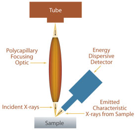
Standard µXRF configuration. The sample can be scanned to measure the elemental distribution within a sample with a spatial resolution as small as 10 µm (energy dependent).
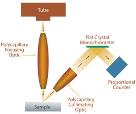
Micro WDXRF instrumentation using a polycapillary focusing optic to focus X-rays from the source to the sample and a polycapillary collimating optic to collect fluorescence emission from a small spot on the sample surface and direct it to the dispersion crystal.
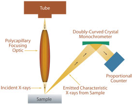
Micro WDXRF using a polycapillary focusing optic to focus X-rays from the source to the sample and a doubly curved crystal monochrometer to collect and disperse fluorescence X-rays from a small spot on the sample and direct them to the detector.
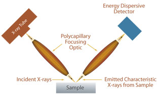
Confocal µXRF uses two polycapillary optics, one for small spot sample excitation and a second to act as a spatial filter for all applications were background radiation from areas not in the region of interest interferes with the signal of interest. This configuration is useful for measurement of spatial distributions in radioactive samples as well as depth profiling applications.
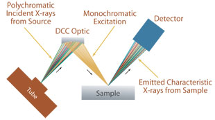
Monochromatic Micro EDXRF using Doubly Curved Crystal Optics.
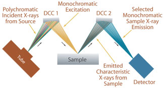
Monochromatic WDXRF using doubly curved crystal optics has the advantage of very high sensitivity for a specific sample element of interest. This technique has been successfully used for measurement of low levels of sulfur in petroleum products.

