-
Analyzers
- Sulfur Analyzers
- Chlorine Analyzers
- Multi Element Analyzers
-
Parts and Supplies
Accessories PartsSupplies
- Service & Maintenance
- Optics & Sources
- Technologies
- Support
- About
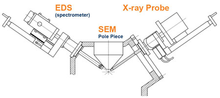
Micro X-Ray Flourescence (??XRF) on Scanning Electron Microscope (SEM)
The capabilities of an SEM can be greatly extended through the addition of an x-ray source with polycapillary optic. The standard Electron Probe X-ray fluorescence (EP-XRF) has some major limitations. First, the electron source causes Bremsstrahlung background which limits the sensitivity of EP-XRF analysis. Secondly, EP-XRF is a surface-sensitive technique and cannot be used to measure underlayer materials in a sample. The integration of an X-ray source to the SEM to perform X-ray excitation XRF allows for much higher detection sensitivity, especially for high-energy X-rays (>5 keV).
Underlayer analysis is possible due to much better depth penetration of X-rays in comparison with the electron probe. Furthermore, XRF analysis allows the sample to be measured “as is”, while a certain level of sample preparation is required for EP-XRF. Also, X-ray excitation allows for operation in air or a low vacuum environment which is critical for bio-chemistry applications. In addition to these benefits a polycapillary optic allows for microfocus spots with high X-ray flux density that provide high resolution and high-speed X-ray mapping.
μXRF on SEM, at a Glance
- Pairing an X-ray source with a polycapillary optic produces a microfocus spot able to provide high resolution and high-speed x-ray mapping
- An X-ray source with optic reduces Bremsstrahlung background radiation, increases sample depth penetration for underlayer analysis, and allows for much higher detection sensitivity
- XRF analysis allows the sample to be measured “as is”, operating in air or low vacuum environments
Product Highlight
A fleX-Beam provides a plug-and-play solution to integrate the µXRF capability into an existing SEM system, extending the functionality of the SEM at lower cost by using existing hardware and software on the SEM. XOS has partnered with iXRF, Inc. to make the technology commercially available.
More details about the commercial products can be found at iXRF website www.ixrfsystems.com.

Features and Benefits
- fleX-Beam’s intensity is up to 10,000 times greater than conventional pinhole collimators
- Focal spot as small as 5µm @ Rh Ka (20.162keV)
- 50-watt performance exceeds conventional kilowatt-powered X-ray tubes
- Integrated safety shutter & 8-position filter wheel
Applications
Micro X-ray Fluorescence (micro-XRF)
- Small Feature Analysis
- Film & Plating Thickness
- High-Resolution Elemental Mapping
- Gun residue study in forensics
- Pigment study in art preservation
- Composition and artifact analysis in archaeology
Talk to an Expert
