-
Analyzers
- Sulfur Analyzers
- Chlorine Analyzers
- Multi Element Analyzers
-
Parts and Supplies
Accessories PartsSupplies
- Service & Maintenance
- Optics & Sources
- Technologies
- Support
- About
Enhancing Micro XRF Analysis with Polycapillary Optics
BACKGROUND
X-ray fluorescence (XRF) is a commonly used technique
for elemental analysis. Compared to other technologies,
XRF is non-destructive, easy to use, and requires
minimal sample preparation. Micro-XRF applies the same
principles as XRF but on a much smaller (micro) scale.
CHALLENGE
To perform micro-XRF, a micron-sized beam of X-rays is required to hit the sample. This small beam can be achieved using a conventional pinhole aperture or X-ray optics. One challenge faced with using a conventional pinhole aperture is that it will block a large portion of the X-rays emitted from the source thereby reducing the number of X-rays hitting the sample, resulting in much lower fluorescence. Another challenge presented is that the output beam from the pinhole will be divergent, resulting in a larger excitation area on the sample. Both challenges impact the detection sensitivity and spatial resolution of micro-XRF analysis.
SOLUTION
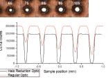
It has been proven that the use of polycapillary focusing optics greatly enhances micro-XRF analysis. Polycapillary optics offer many benefits; a large collection solid angle, high flux density, and a focused micron sized beam. Polycapillary optics transmit a polychromatic beam that can achieve a focused spot size as small as 5μm, FWHM at Rh Kα energy (20.2keV) with intensities greater than 107 photons/second. These performance attributes provide a great impact to micro-XRF across many different applications. Figure 1 below is an example of a mapping application.
Figure 1
1D XRF scan (bottom), of a copper PCB sample (top), using both a regular and a halo reduction polycapillary optic, each with a 15μm focal spot. The high-energy halo effect is clearly visible with regular optic while it is eliminated with the halo reduction optic.
1D XRF scan (bottom), of a copper PCB sample (top), using both a regular and a halo reduction polycapillary optic, each with a 15μm focal spot. The high-energy halo effect is clearly visible with regular optic while it is eliminated with the halo reduction optic.
CONCLUSION
The use of polycapillary optics has completely changed the analytical speed and accuracy micro-XRF analysis. Accurate and reliable measurement results can be achieved in hours compared to a conventional approach which may take days. This technique benefits many applications such as plating thickness, elemental mapping, forensics and cultural heritage. XOS offers customized polycapillary optic solutions optimized for your application needs, as well as a comprehensive solution for OEM or lab-based systems known as fleX-Beam™.
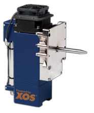
fleX-Beam™ is a unique, compact X-ray generator that combines a low-powered X-ray source and a precisely-aligned polycapillary optic to deliver a bright X-ray beam for advanced material analysis. fleX-Beam is available in several standard focused or collimated beam configurations and can also be customized for specific applications. This is the ideal solution for any OEM or lab-based system.