-
Analyzers
- Sulfur Analyzers
- Chlorine Analyzers
- Multi Element Analyzers
-
Parts and Supplies
Accessories PartsSupplies
- Service & Maintenance
- Optics & Sources
- Technologies
- Support
- About
X-Ray Fluorescence in Scanning Electron Microscopy (SEM)
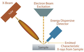
Electron-probe X-ray fluorescence (EP-XRF) analysis has been widely used in scanning electron microscopy instrumentation for elemental analysis of specimens. With the x-ray fluorescence microscopy in the SEM, the surface of a solid sample is excited with a highly-focused energetic beam of electrons which induces X-ray fluorescence from the elements within the sample.
The major advantage offered by the electron excitation, as opposed to X-ray excitation, is the small probe size, typically < 1 ?m, and its high excitation efficiency, especially in the low-energy range. However, EP-XRF has some major limitations. First, the electron source causes Bremsstrahlung background which limits the sensitivity of EPXRF analysis. Secondly, EPXRF is a surface-sensitive technique and cannot be used to measure underlayer materials in a sample. In contrast, XRF analysis using X-ray excitation offers much higher detection sensitivity, especially for high-energy X-rays. Underlayer analysis is possible due to much better depth penetration of X-rays in comparison with the electron probe. Furthermore, XRF analysis allows the sample to be measured “as is”, while a certain level of sample preparation is required for EPXRF, as it requires that sample is conductive.
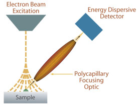
The capability of excited X-ray fluorescence can be integrated into an existing SEM system using an X-Beam unit containing a polycapillary optic. The approach extends the capability of the SEM at low cost by using existing hardware and software on the SEM.